Naoya Kurata

and 12 more

Background: The efficacy of ablation targeting low-voltage areas (LVAs) is controversial, although LVA presence is well known to be associated with AF recurrence after ablation. Atrial fibrillation (AF) substrate may not localize within LVAs. Methods and results: This observational study enrolled 405 consecutive patients who underwent an initial AF ablation procedure. The left atrial voltage map was obtained after pulmonary vein isolation. LVAs were defined as areas with voltage < 0.5 mV. To estimate whole atrial electrophysiological degeneration, mean regional voltage at each of 6 regions and left atrial total conduction velocity were measured. LVAs existed in 143 of 405 (35.3%) patients. Patients with LVAs demonstrated lower mean regional voltages throughout all 6 regions than those without LVAs (1.3 [1.8, 0.8] vs. 0.6 [1.0, 0.2] for anterior wall, p<0.001). On the other hand, left atrial conduction velocity was lower in patients with LVAs than in those without (0.89 [1.01, 0.74] vs. 0.93 [1.03, 0.87] m/s, p<0.001). Multivariate analysis revealed that low left atrial total conduction velocity and a higher number of regions with mean voltage reduction were independently associated with AF recurrence, although LVA presence was not. Conclusion: Patients with localized left atrial LVAs were characterized by whole left atrial electrophysiological degeneration as assessed by mean regional voltage and conduction velocity. In addition, whole left atrial electrophysiological degeneration parameters were well associated with AF recurrence.

Masaharu Masuda

and 12 more

Introduction: A novel ablation catheter that can measure local impedance (LI) was recently launched. We aimed to explore target LI measurements at each radiofrequency application (RFA) for creating sufficient ablation lesions during pulmonary vein (PV) isolation. Methods: This prospective study included 15 consecutive patients scheduled to undergo an initial ablation of paroxysmal atrial fibrillation (AF). Circumferential ablation around both ipsilateral PVs was performed using a 4-mm irrigated ablation catheter with an LI sensor. Point-by-point ablation was used with a 4-mm inter-ablation-point distance. Operators were blinded to LI measurements during the procedure. Creation of sufficient ablation lesions was assessed by the absence of a conduction gap. Results: After first-pass encircling PV antrum ablation, left atrium to PV conduction remained in 12 of 30 (40%) ipsilateral PVs. Mapping using the mini-basket catheter identified 48 ablation points through which the propagation wave entered the PV. At ablation points with a gap, the LI drop during RFA was half that at points without a gap (12 ± 7 vs. 23 ± 12 ohm, p<0.001). The GI drop did not differ between ablation points with and without a gap (12 ± 7 vs. 14 ± 10 ohm, p=0.10). An LI drop of 15 ohm predicted sufficient lesion formation without a gap with a sensitivity of 0.71, specificity of 0.81, and predictive accuracy of 0.75. Conclusion: A target LI drop of 15 ohm at each RFA with a 4-mm distance between adjacent ablation points may facilitate creation of sufficient ablation lesions during PV isolation

Naoya Kurata

and 11 more

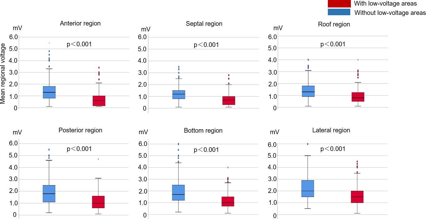
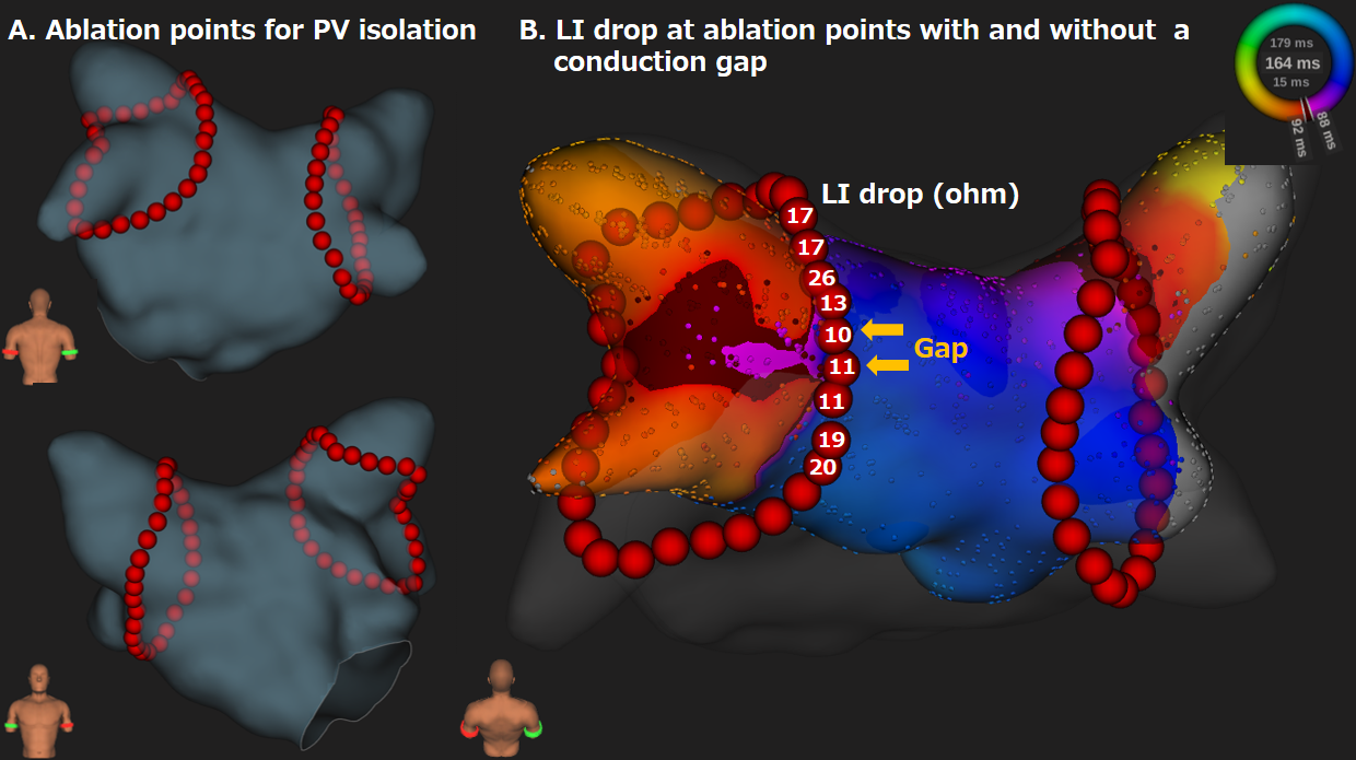
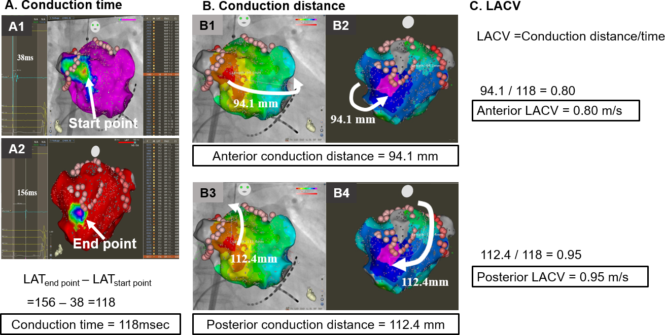
Background: Atrial conduction velocity may represent atrial fibrillation (AF) substrate after pulmonary vein isolation (PVI). To elucidate the association between whole left atrial conduction velocity (LACV) and AF recurrence after PVI. Methods and Results: This observational study enrolled 279 patients who underwent PVI alone as an initial AF ablation procedure. After PVI, the left atrium was mapped with a 20-pole multielectrode in conjunction with the CARTO3 system during 100-ppm right atrial pacing. Left atrial conduction distance and conduction time were calculated from the start to the end of the propagation wave front in the left atrium. LACVs on the anterior and posterior routes were calculated as conduction distance divided by conduction time. Anterior and posterior LACVs were slower in patients with AF recurrence than in those without (anterior, 0.79 [0.71, 0.86] vs. 0.96 [0.90, 1.06], p < 0.001; posterior, 0.99 [0.89, 1.14] vs. 1.10 [1.00, 1.29], p < 0.001). AF recurrence was best predicted by anterior LACV with a cut-off value of 0.87 m/s (sensitivity 87%, specificity 81%, and predictive accuracy 84%). Multivariate analysis demonstrated that a slow anterior LACV < 0.87 m/s was an independent predictor of AF recurrence with an adjusted hazard ratio of 11.8 (6.36 – 22.0). Patients with anterior low-voltage areas demonstrated slower anterior LACV than those without low-voltage areas (0.89 [0.71, 1.00] vs. 0.94 [0.87, 1.05], p < 0.001). Conclusion: A slow LACV in the entire left atrium was an excellent predictor of AF recurrence after PVI, suggesting the necessity of additional ablations.