Masato Hachisuka

and 9 more

Introduction: Atrial fibrillation (AF) is the most common arrhythmia in patients undergoing hemodialysis (HD), which lowers the quality of life (QoL) and increases the risk of dialysis related complications. The present study aimed to evaluate the effectiveness of AF ablation on the QoL in patients undergoing HD. Methods and Results: Nineteen patients undergoing HD (14 men, age 68±8years, 15 paroxysmal AF) who underwent catheter ablation (CA) of drug-refractory AF were enrolled in the study. The ablation outcomes and procedural complications were evaluated and compared to 1053 consecutive patients without HD who underwent AF ablation. The Kidney Disease Quality of Life Short Form (KDQOL-SF) was assessed to evaluate the QoL of the HD patients at baseline and six months after the ablation. During the follow-up period of 17±13 months after the last procedure, the arrhythmia free rate was similar (HD patients 79% vs. non-HD patients 86%, log-rank p=0.82). There were no life-threatening complications in any patients. The KDQOL-SF of the HD patients six months after the ablation showed an improvement in the physical functioning (54±23 to 68±28, p<0.01), general health perceptions (38±17 to 48±15, p<0.01) and symptoms/problems (75±21 to 84±13, p=0.02) as compared to the baseline. Regarding the intradialytic symptoms, the dyspnea during HD significantly improved after the CA in the HD patients without AF recurrence (35% to 6%, p=0.04), while the atrial tachyarrhythmias and hypotension during HD remained unchanged. Conclusions: CA of AF improves the QoL in patients with chronic hemodialysis.

Rei Mimuro

and 9 more

Introduction: Atrial fibrillation (AF) is a common arrhythmia in patients with hypertrophic cardiomyopathy (HCM) and is associated with renal function deterioration. The protective effects of catheter ablation (CA) of AF on the renal function in HCM patients remains unsolved. Methods: From 2009 to 2020, 169 consecutive patients with HCM and AF (age 70±12, 87 males) were retrospectively evaluated. The estimated glomerular filtration rate (eGFR) was evaluated at the study enrollment or one month before the CA and reevaluated three months and 12 months later. Results: Among the 169 patients, 63 underwent CA of AF (ablation group) and the remaining 106 did not (control group). After propensity score matching, 45 pairs were matched. The baseline eGFR was similar between the two groups (P=0.83). During a mean follow-up period of 34±27 months, sinus rhythm was maintained in 36 (80%) patients after 1.7±0.8 ablation procedures. The eGFR significantly decreased from baseline to three months (P<0.01) and from baseline to one year (P<0.01) in the control group, while the eGFR in the ablation group was maintained both from baseline to three months (P=0.94) and from baseline to one year (P=1.00) after the CA. The change in the eGFR between baseline and 12 months was significantly smaller in the ablation group than control group (P<0.01). After a logistic regression analysis, CA of AF was the independent predictor of an improvement of eGFR (OR: 2.81; 95% CI: 1.08-7.36 P=0.04). Conclusions: CA of AF had a protective effect on the renal function in patients with HCM.

Kanako Ito-Hagiwara

and 9 more

Background: Atrial flutter (AFL) is a large re-entrant circuit located in the right atrium. Anti-arrhythmic drugs (AADs) can provoke AFL with 1:1 atrioventricular conduction (AVC) to cause hemodynamic collapse. We elucidated the characteristics of patients with AFL exhibiting spontaneous 1:1 AVC. Methods: Fifteen patients (1:1 AFL group; 11 males, 52.4±13.7 years old) who documented AFL with 1:1 AVC were enrolled and compared to 77 patients without 1:1 AVC (Control group; 71 males, 68.1±10.9 years old). Results: The use of AADs was greater in the 1:1 AFL group than in the control group (60.0 vs. 14.3%, p < 0.001). AFL cycle length during maximum AVC was significantly longer in the 1:1 AFL group than in the control group (274.7 ± 37.0 vs. 220.4 ± 26.2 msec, p < 0.001). Among 1:1 AVC group, 9 patients had AADs and AFL cycle length was significantly longer during 1:1 AVC as compared with 2:1 AVC documented the other day (284.4 ± 41.3 vs. 233.3 ± 26.0 msec, p <0.001), suggesting enhancement effect of the AADs during 1:1 AVC. Remaining 6 patients who did not take AADs, 2 patients showed enlargement of the tricuspid annulus and 3 patients developed 1:1 AVC during exercise. Conclusions: In addition to the enhancement of AAD effect, prolonged AFL cycle length associated with enlargement of the tricuspid annulus and shortened refractory period of the AV node might increase the risk of 1:1 AVC during AFL. Keywords: atrial flutter, atrial flutter cycle length, tricuspid annulus. Atrioventricular node, atrioventricular conduction, anti-arrhythmic drug

Kenji Yodogawa

and 7 more

Background The clinical course and therapeutic strategies in the congenital long QT syndrome (LQTS) are genotype-specific. However, accurate estimation of LQTS-genotype is often difficult from the standard 12-lead ECG. Objectives This study aims to evaluate the utility of QT/RR slope analysis by the 24-hour Holter monitoring for differential diagnosis of LQTS-genotype between LQT1 and LQT2. Methods This cross-sectional study enrolled 54 genetically identified LQTS patients (29 LQT1 and 25 LQT2) recruited from 3 medical institutions. The QT-apex (QTa) interval and the QT-end (QTe) interval at each 15-second were plotted against the R-R intervals and the linear regression (QTa/RR and QTe/RR slopes, respectively) were calculated from the entire 24-hour and separately during the day or night-time periods of the Holter recordings. Results The QTe/RR and QTa/RR slopes at the entire 24-hour were significantly steeper in LQT2 compared to those in LQT1 patients (0.262 +/- 0.063 vs 0.204 +/- 0.055, P = 0.0007; 0.233 +/- 0.052 vs 0.181 +/- 0.040, P = 0.0002, respectively). The QTe interval was significantly longer, QTe/RR and QTa/RR slopes at daytime were significantly steeper in LQT2 than in LQT1 patients. The receiver operating curve analysis revealed that the QTa/RR slope of 0.211 at the entire 24-hour Holter was the best cut-off value for differential diagnosis between LQT1 and LQT2 (sensitivity: 80.0%, specificity: 75.0% and area under curve: 0.804 [95%CI = 0.68-0.93]). Conclusion The continuous 24-hour QT/RR analysis using the Holter monitoring may be useful to predict the genotype of congenital LQTS, particularly for LQT1 and LQT2.

Ippei Tsuboi

and 9 more

Introduction: There is increasing evidence of the epicardial connection between the right-sided pulmonary vein (PV) carina and right atrium interrupts right-sided PV isolation after circumferential PV ablation in patients with atrial fibrillation. In such cases, carina ablation is often required. This study aimed to assess the utility of the right atrial posterior wall (RAPW) pacing in the detection of the right-sided epicardial connection (EC), evaluate the requirement for additional carina ablation after circumferential pulmonary vein (PV) ablation depending on the presence of EC, and investigate the clinical characteristics including the amount of epicardial adipose tissue (EAT) in patients with ECs. Methods and Results: Forty-one patients scheduled for PV isolation were enrolled. Before ablation, activation mapping of the LA was prospectively performed during pacing from the RAPW. EC was observed in 12 patients (EC group, 29%), whereas no EC was observed in the remaining 29 patients (non-EC group, 71%). For PV isolation, carina ablation was required in addition to circumferential ablation in 7 patients (58%) in the EC group, compared to 2 patients (7%) in the non-EC group (p < 0.003). Periatrial and intercaval EAT volumes were significantly lower (12.8 ± 6.2 vs. 23.1 ± 13.9 ml/m 2, p < 0.02, and 1.1 ± 0.8 vs. 2.2 ± 1.6 ml/m 2, p< 0.02, respectively) and the patients were younger (66.5 ± 6.6 vs. 72.4 ± 8.3 years, p < 0.03) in the EC group than in the non-EC group. Conclusions: RAPW pacing revealed EC between the RA and right PV carina in nearly a quarter of the patients.

Keisuke Usuda

and 34 more

Michio Ogano

and 4 more

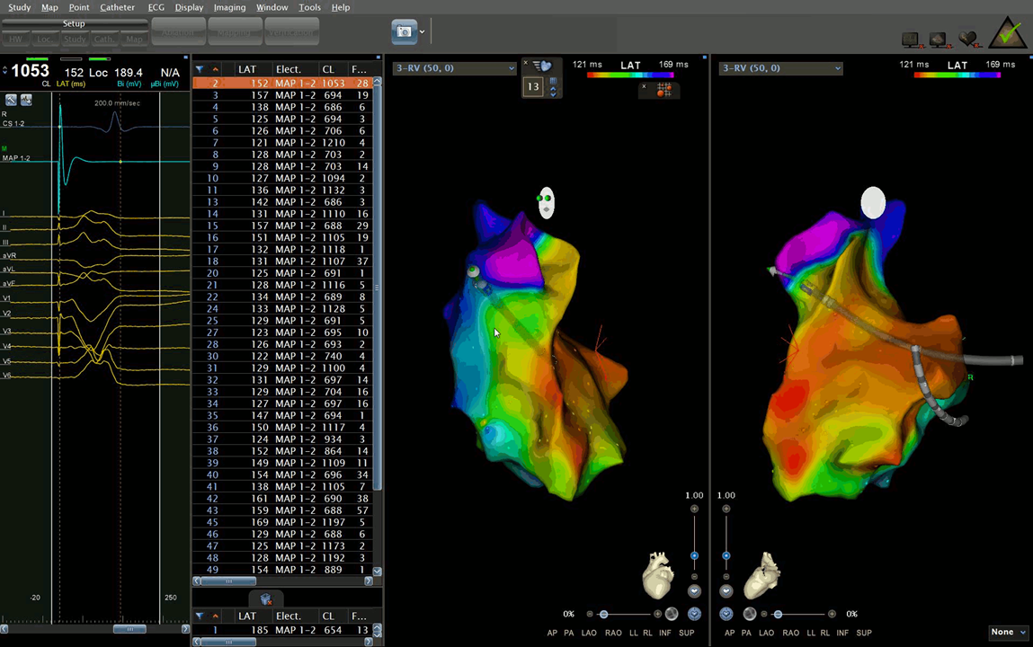
Introduction: Right ventricular (RV) pacing causes changes in the heart’s electrical and mechanical activation patterns. QRS duration is a useful surrogate marker of electrical dyssynchrony; longer QRS duration during RV pacing indicates poor prognosis. However, the mechanisms underlying longer QRS duration during RV pacing remain unclear; hence, we investigated factors predicting QRS prolongation during RV pacing. Methods and Results: We enrolled 211 patients who underwent catheter ablation for supraventricular tachyarrhythmia and showed no bundle-branch-block. Three-dimensional mapping for QRS duration during RV pacing from the RV outflow to RV apex was performed, and the difference in QRS duration was analyzed. The predisposing factors causing QRS >160 ms during RV apical pacing were also analyzed. QRS durations at baseline and during RV pacing from the RV outflow and at RV apex were 85.0±7.5 ms, 163.7±17.1 ms, and 156.2±16.1 ms, respectively. With respect to QRS duration, there was a significant correlation between RV outflow and RV apical pacing (r=0.658, p<0.001). The difference in QRS duration between RV outflow and apex in each patient was only 12.5±10.4 ms. Logistic multivariable regression analysis identified baseline QRS duration [odds ratio (OR) 1.24, 95% confidence interval (CI) 1.15 to 1.33, p<0.01], interventricular septum thickness (OR 1.20, 95% CI 1.02-1.40, p=0.025), left atrial diameter (OR 1.08, 95% CI 1.01-1.16, p=0.024), and E/e’ (OR 1.23, 95% CI 1.12-1.35, p<0.01) as significant predictors of prolonged QRS duration during RV apical pacing. Conclusion: QRS duration during RV pacing largely depends not on the pacing site, but on underlying structural heart diseases.