Alessandro Barbone

and 10 more

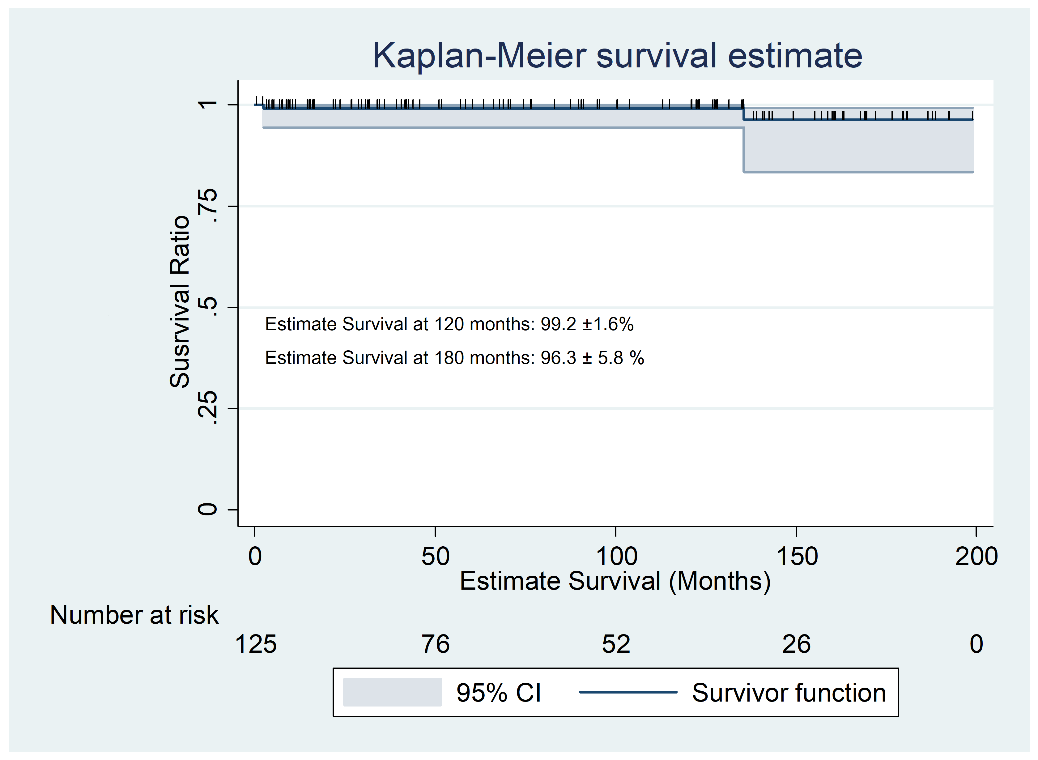
Background and Aim of the Study: Barlow’s disease is characterized by excess myxomatous degenerative tissue, leaflets prolapse and/or billowing, chordal-elongation, and annular-dilation. We reviewed our experience in Minimally Invasive Mitral Valve Repair (MIMVR) techniques to treat this complex mitral pathology. Methods: Between 1999 and 2017 a group of 125 patients was identified as Barlow’s disease undergoing MIMVR. The diagnosis of Barlow disease was based upon preoperative transesophageal-echocardiography (TEE) and confirmed by the surgeon’s assessment during open-heart procedure. Operative technique and data were retrospectively collected along with intra-hospital and long-term follow-up. Results: Successful Mitral Repair was possible in 100% of cases (125 patents) within the first cross clamp. Most patients (118 – 94.4%) were treated by the Edge to Edge (Alfieri-Stich), while 2 (1.6%) where corrected by neochordae implant and 2 (1.6%) by quadrangular resection. 58 patients (47.1%) received complete semi-rigid ring, while 65 (52.9%) incomplete flexible partial ring excluding the LAM. Concomitant procedures were additional cleft occlusion in 10 cases (8%), tricuspid valve repair (ring implant or remodeling) in 29 cases (23.2%), left atrial ablation for atrial fibrillation in 4 cases (3.2%) and atrial septal repair in 4 (3.2%). Operative mortality was 0%. Average long-term follow-up was 85 ± 62 months, with a survival rate of 97.6%, freedom from redo mitral surgery of 98.4% and freedom from >2+ recurrent mitral regurgitation of 94.5%. No difference in outcome could be related to annuloplasty ring type. Conclusions: Mitral repair can be safely and successfully achieved though minimal-invasive approach, with optimal long-term results.

Alessandro Barbone

and 3 more

Paragangliomas (PGLs) are rare neuroendocrine tumors that arise from the autonomic ganglia. A 34-year-old woman was admitted to our department with palpitation, fatigue and chest pain under stress. Past medical history included a para-carotid PGL surgically removed 5 years earlier. Her family history was notable for familiar PGL with SDH mutation. At stress-echocardiography, was recorded a marked ST-elevation on precordial-leads, concomitant to new onset anterior septum hypo-kinesis, and therefore the patient was proposed for coronary angiography. Although angiographic evidence of atherosclerosis were not recorded, was found that Left Anterior Descending Coronary Artery gave off some branches towards a vascular structure (3×2 cm) in contiguity with the left coronary sinus of the aortic root. Subsequent Compute-Tomography and Magnetic-Resonance clarified the anatomical relationships of the mass with adjacent cardiac structures, in particular the mass was located behind the pulmonary artery trunk and lateral to aortic root, lying above the first portion of the anterior descending coronary artery. Subsequently a careful discussion about the risks and benefits of all therapeutic opportunity, the open surgical resection was deemed the best option for the patient despite the complex clinical anatomy. After standard cannulation for CPB and cardioplegic arrest, transection of the pulmonary artery and the aorta was performed granting adequate exposure of the mass and identification of afferent and efferent vascularization for complete resection though a cleavage plane. To ensure adequate hemostasis on a such critical area, the continuity of the pulmonary trunk was ensured only after removing aortic cross-clamp. The post-operative course was uneventful