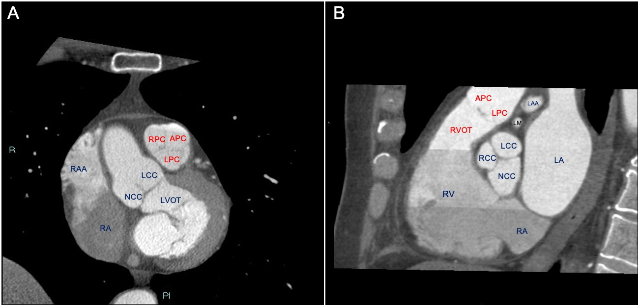
Ventricular tachycardia and premature ventricular complexes (PVCs) arising from right ventricular outflow tract (RVOT) are the most common type of ventricular arrhythmias (VAs) in patients without structural heart disease. Radiofrequency ablation is now the gold standard of treatment in this setting due to high efficacy rates and optimal safety profile [2] During the last few years, the pulmonary valve (PV) and the pulmonary artery (PA) have attracted much attention as reliable sites of origin of RVOT-type arrhythmias. In the mean while intracardiac echocardiogram (ICE) has undoubtedly improved our understanding and approach to manage these arrhythmias accurately characterizing the PV and its contiguous structures. Aim of this paper is to provide an illustrated step-by-step guide on how to use ICE with the CARTOSOUND module to visualize and reconstruct 3D shell of the RV, the PV, as well of other anatomical structures (i.e., the aortic valve and coronary arteries) to perform aware and safe ablation in this region. A new reconsideration of the existent classification of these VAs is also provided.