Essential Maintenance: All Authorea-powered sites will be offline 9am-10am EDT Tuesday 28 May
and 11pm-1am EDT Tuesday 28-Wednesday 29 May. We apologise for any inconvenience.

Background:This prospective study aims to reveal whether the lesion is a benign pathological process or malignant by measuring ADC values under PI-RADSv2.1 guidance on MpMRI examinations. Additionally, the paper evaluates whether there is a correlation between malignant lesions’ pathological grade and ADC values, and whether ADC values provide noninvasive information about prostate cancer aggressiveness. Purpose:To determine the cut-off ADC values that may exist to identify and distinguish between benign and malignant lesions and also identify cancers with an ISUP score≥2 and cancers with an ISUP score1 defined as silent disease. Methods:This study includes 243 patients and they were diagnosed with TRUS-guided cognitive MRI fusion as tissue diagnosis. MpMRI images were evaluated before biopsy according to PI-RADSv2.1 guideline by a radiologist. Three groups which are benign prostatic tissue, prostatitis, prostate cancer, were obtained according to the histopathological results. Results: When the cut-off value for ADC is 780 x10-3, sensitivity was 80%. When the cut-off value was taken as 668 x10-3, the sensitivity was found to be 72% and specificity 62%. When the cut-off ADC value was taken as 647 x10-3, the sensitivity was 83% and the specificity was 48.5%. ADC values varied significantly according to ISUP groups [p= 0.003]. It was determined that ISUP 1 group was significantly higher than each of the other groups. ADC group mean values did not show a statistically significant difference between Group 2,3,4 and 5. Conclusion:ADC value shows significant potential, and may it improve the diagnostic accuracy.

Ali Murat Koc

and 4 more

Aims Delay and false positivity in PCR test results have necessitated accurate chest CT reporting for management of patients with COVID-19 suspected symptoms. Pandemic related workload and level of experience on covid-dedicated chest CT scans might have effected diagnostic performance of on-call radiologists. The aim of this study is to reveal the interpretation errors in chest-CT reports of COVID-19 suspected patients admitted to the ER. Methods COVID-19 dedicated chest-CT scans which were performed between March and June 2020 were re-evaluated and compared with the former reports of these scans and PCR test results. CT scan results were classified into four groups. Parenchymal involvement ratios, radiology departments’ workload, COVID-19 related educational activities have examined. Results Out of 5721 Chest-CT scans, 783 CTs belonging to 664 patients (340 female, 324 male) were included to this study. RT-PCR test was positive in 398; negative in 385 cases. PCR positivity was found to be highest in “normal” and “typical for covid” groups whereas lowest in “atypical for covid” and “not covid” groups. 5-25% parenchymal involvement ratio was found in 84.2% of the cases. Regarding number of chest CT scans performed, radiologists’ workload have found to be increased six-folds compared to the same months of the former year. With the re-evaluation, a total of 145 IEs (18.5%) have been found. IEs were mostly precipitated in the first two months (88.3%) and mostly in “not covid” class (60%) regardless of PCR positivity. COVID-19 and radiology entitled educational activities along with the ER admission rates within the first two months of pandemic have seem to be related with the decline of IEs within time. Conclusion COVID-19 pandemic made a great impact on radiology departments with an inevitable burden of daily chest-CT reporting. This workload and concomitant factors have possible effects on diagnostic challenges in COVID-19 pneumonia.
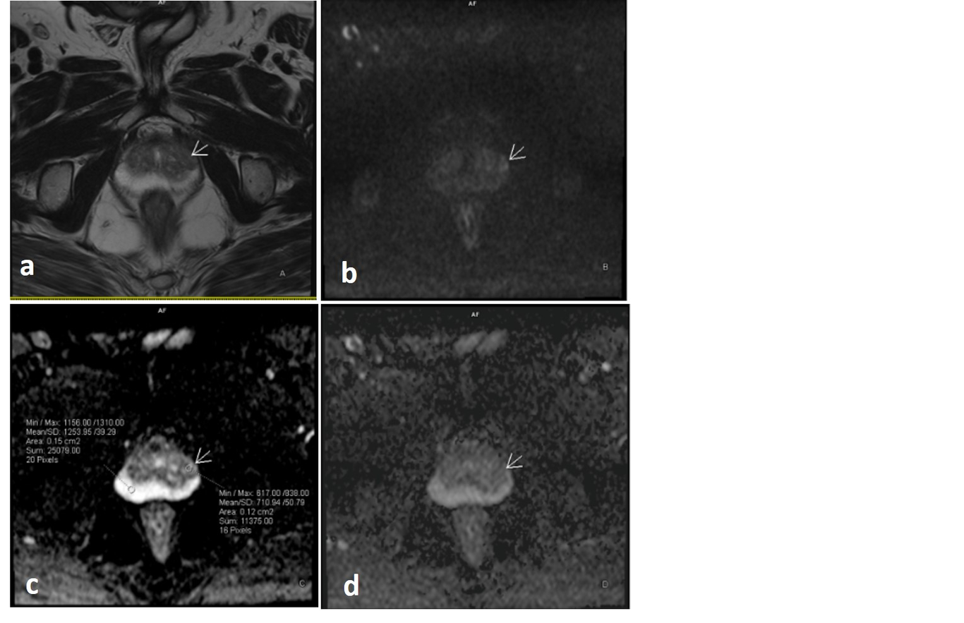
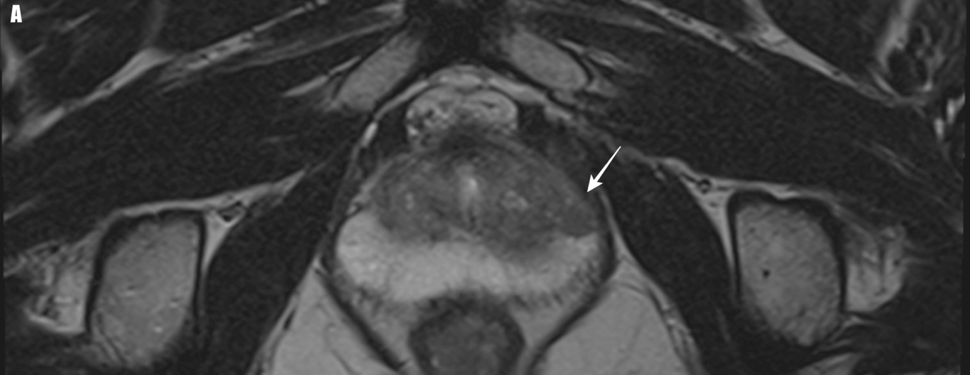
Purpose: To evaluate the correlation between PI-RADSv2.1 and International Society of Urologic Pathologists (ISUP) score for patients who underwent multiparametric-MRI(MpMRI) prior to transrectal ultrasound (TRUS) guided cognitive fusion biopsy (CF-Bx). And to investigate inter-observer agreement of PI-RADSv2.1. Methods: Patients who underwent MpMRI of prostate prior to first TRUS-guided CF-Bx, were included in this prospective study. MpMRI examinations were evaluated by two radiologists before biopsy according to the PI-RADSv2.1. Interobserver agreement was recorded and the final PI-RADS categorization was performed by consensus. Correlation of histopathological results with PI-RADSv2.1 score was evaluated. Lesions with Gleason Score(GS)≥6 were considered as prostate cancer (PCa). Results: A total of 84 patients with 106 lesions were included in the study. The ratio of PCa in the PI-RADS groups 1,2,3,4,5 was 0%, 0%, 22.2%, 56%, 94.45%, respectively. There was a positive correlation with a value of 0.814 between the PI-RADSv2.1 and the ISUP score. When PI-RADS≥3 is accepted as the cut-off value in peripheral zone(PZ) and the whole gland, the NPV for malignancy was 100.00%. For PI-RADS ≥4, it was 76.47% for PZ, and 80.65% for the whole gland. For the whole gland; sensitivity, specificity, and PPV of the PI-RADS≥3 were 100%, 12.9%, and 44.33%, respectively; for PI-RADS≥4, these values were 72.09%, 80.65%, and 72.09% respectively. Without applying cut-off values, the interobserver agreement for PI-RADS score was κappa:0.562. Conclusions: PI-RADSv2.1 was created in the framework of v2 to facilitate to evaluate MpMRI and to increase interobserver agreement. We believe that further studies will be necessary.