Jakub Holda

and 8 more

Introduction: The aim of this study was to describe the morphology of the right atrioventricular valve (RAV) and determine its spatial position in relation to selected structures of the right atrium. Methods and Results: We examined 200 randomly selected human adult hearts. All leaflets and commissures were identified and measured. The position of the RAV was defined. 3-leaflet configurations were present in 67.0% of cases whereas 4-leaflet configurations were present in 33.0%. Valves with four leaflets have significantly larger perimeter (119.2±11.1 vs. 109.3±11.3mm, p=0.001). No significant difference was found in superior leaflet length and height between 3- and 4-leaflet RAVs. Septal and mural leaflets were both significantly shorter and higher in 4-leaflet than in 3-leaflet RAVs. Significant domination of the muro-septal commissure in 3-leflet valves was noted. The supero-septal commissure was the most stable point within RAV circumference, with no difference in its position between 3- and 4-leaflet valves. In 3-leaflet valves the muro-septal commissure was placed within cavo-tricuspid isthmus area in 52.2% of cases, followed by the right atrial appendage vestibule region (20.9%). In 4-leaflet RAVs, the infero-septal commissure was located predominantly in the cavo-tricuspid isthmus area and infero-mural commissure was always located within the right atrial appendage vestibule region. Conclusions: The RAV is a highly variable structure. The supero-septal part of the RAV is the least variable component, whereas the infero-mural is the most variable. The number of detected RAV leaflets significantly influences the relative position of individual valve components in relation to right atrial structures.

Michał Bonczar

and 9 more

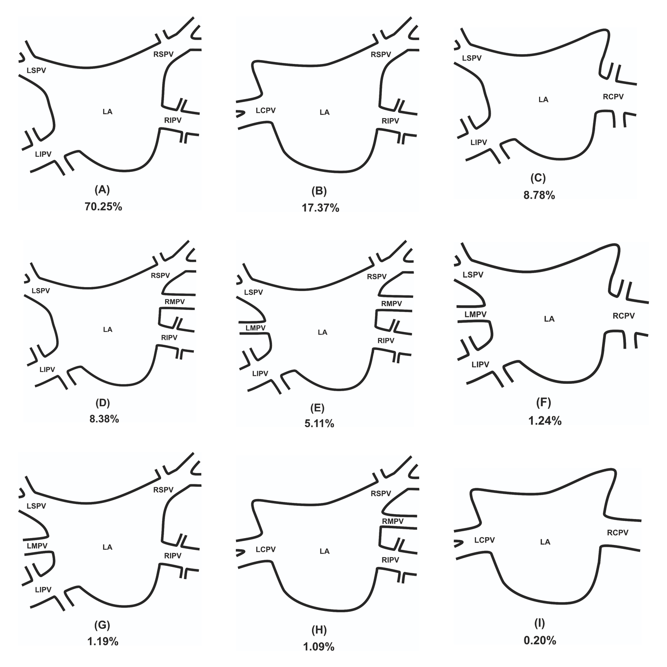
Introduction: The aim of this study was to establish the most accurate and up-to-date anatomical knowledge of PVs ostia variations, diameters and ostial area, in order to provide physicians, especially heart and thoracic surgeons with exact knowledge concerning this area. Materials and methods: Major online medical databases such as PubMed, Embase, Scopus, Web of Science and Google Scholar were searched to gather all studies in which the variations, maximal diameter and ostial area of the PVs were investigated. During the study, the Preferred Reporting Items for Systematic Reviews and Meta-Analyses (PRISMA) guidelines were followed. Additionally, The Critical Appraisal Tool for Anatomical Meta-analysis (CATAM) was used to provide the highest quality findings. Reults: The most common ostia variation is classical one, containing left superior pulmonary vein (LSPV), left inferior pulmonary vein (LIPV), right superior pulmonary vein (RSPV) and right inferior pulmonary vein (RIPV). Mean diameters and ostial areas of each pulmonary vein were established in general population and in multiple variations considering method of gathering the data and geographical location. Conclusion: Significant variability in PV ostia is observed. Left-sided PVs have smaller ostia than corresponding right-sided PVs, and the inferior PVs ostia are smaller than superior. The size of the LCPV ostium is the largest among all analyzed veins, while the ostium of RMPV is the smallest. It is hoped that the results from this meta-analysis will help clinicians in planning and performing procedures that involve pulmonary and cardiac area.

Jakub Holda

and 7 more

Introduction: The aim of our study was to investigate the presence and mutual relationships of coronary vessels within the right atrial appendage RAA vestibule. Methods and Results: We examined 200 autopsied hearts. The RAA vestibule was cross sectioned along its isthmuses (superior, middle, and inferior). We assessed the presence and mutual relationships between coronary blood vessels. The right coronary artery (RCA) was present in 100% of the superior RAA isthmuses but absent in 2.0% of hearts within the middle isthmus and in 6.5% of hearts within the inferior RAA isthmus. Its diameter was quite uniform along the superior (2.6±0.8mm), middle (2.9±1.1mm) and inferior (2.7±0.9mm) isthmuses (p=0.12). The location of the RCA varied significantly, and it was sometimes accompanied by other accessory coronary vessels. In all the isthmuses, the RCA ran significantly closer to the endocardial surface than to the epicardial surface (p<0.001). At the superior RAA isthmus, the artery was furthest from the right atrial endocardial surface and this distance gradually decreased between the middle RAA isthmus and the inferior RAA isthmus (9.0±4.0 vs. 6.2±3.0 vs. 4.8±2.3mm, respectively; p<0.001). The interposed RCA was found in 7.0% of cases within the superior isthmus, in 2.5% within the middle isthmus and in 1.5% within the inferior isthmus. Conclusions: This study was the most complex analysis of the mutual arrangements and morphometric characteristics of coronary blood vessels within the RAA vestibule. Awareness of additional blood vessels within the vestibule can help clinicians plan and perform safe and efficacious procedures in this region.