Yuji Motoike

and 8 more

Background: Ablation index (AI) linearly correlates with lesion depth and may yield better therapeutic performance in pulmonary vein isolation (PVI) when tailored to a patient’s wall thickness (WT) in the left atrium (LA). Methods and results: (First study) In paroxysmal atrial fibrillation patients (PAF, n=20), the average LA WT (mm) in each anatomical segment for PVI was measured by intra-cardiac echocardiography (ICE) placed in the LA; the optimal AI for creating one-millimeter transmural lesion (AI/mm) was calculated. (Second study) PAF (n=80) patients were randomly assigned either to a force-time integral protocol (FTI, 400 gram·second, n=40) or a tailored-AI protocol (TAI, n=40). In TAI, the LA WT in each segment was individually measured by ICE before starting ablation; a target AI was adjusted according to the individual WT in each segment (AI/mm×WT). The acute procedure outcomes and the 1-year AF recurrence rate were compared between FTI and TAI. TAI had higher success rate of first-pass isolation and had lower incidence of residual PV-potentials/conduction gaps after a circular ablation than FTI (88% vs. 65%, 15 vs. 45%, respectively). The procedure time to complete PVI decreased in TAI compared to FTI (52 vs. 83 minutes), being attributed to the increased radiofrequency power and the decreased radiofrequency application time in each point in TAI. TAI had lower 1-year AF recurrence rate than FTI. Conclusion: WT-based AI-adjustment increased acute procedure success, decreased time for PVI, and reduced 1-year AF recurrence rate. Understanding the precise ablation target would improve the efficacy of PVI.

Masahide Harada

and 9 more

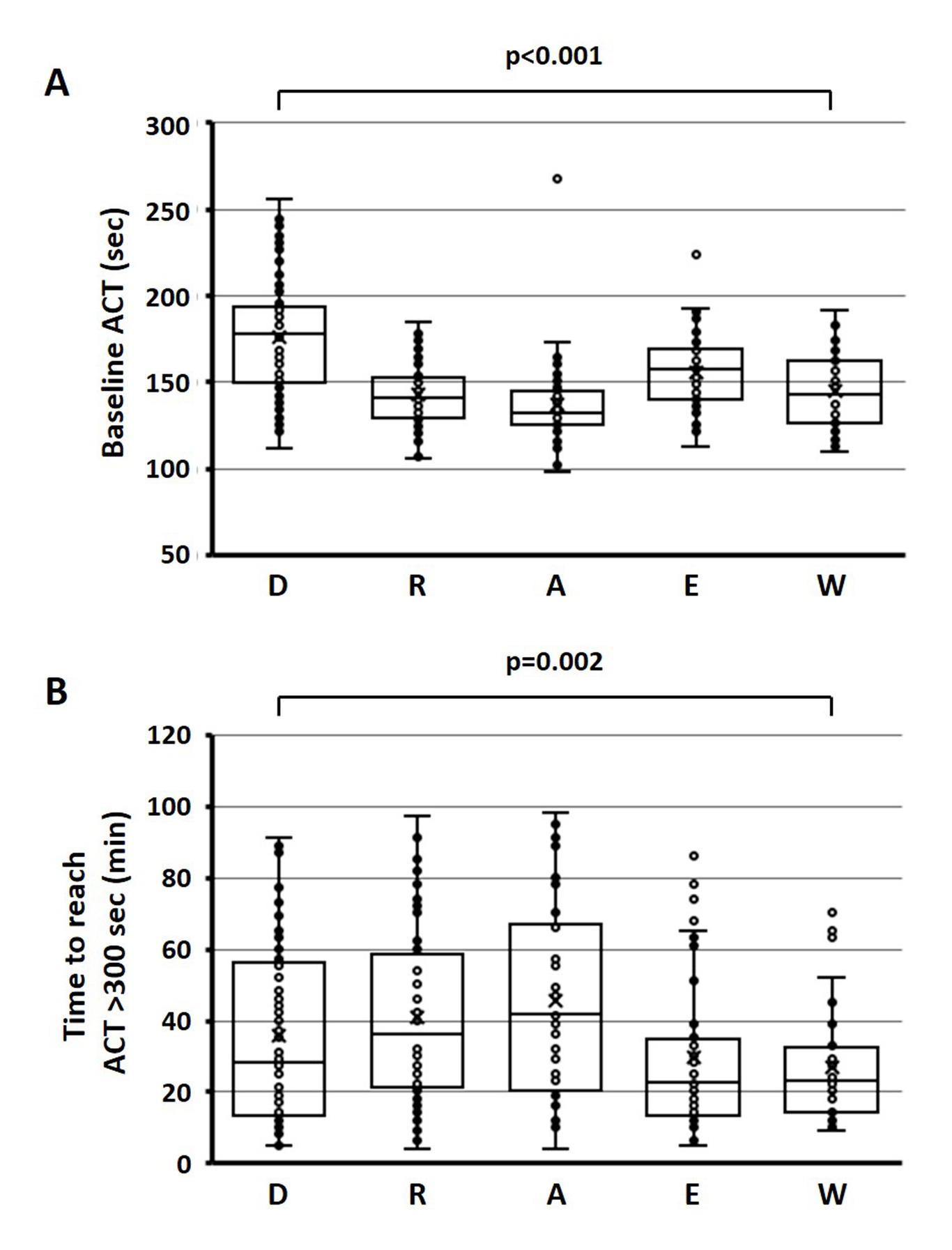
Introduction: Silent cerebral events (SCEs) are related to the potential thromboembolic risk in atrial fibrillation (AF) ablation. Peri-procedural uninterrupted oral anticoagulation (OAC) reportedly reduced the risk of SCEs, but the incidence still remains. Methods and Results: AF patients undergoing catheter ablation were eligible. All patients took non-vitamin K antagonist oral anticoagulants (NOACs, n=248) or vitamin K antagonist (VKA, n=37) for peri-procedural OAC (>4 weeks) without interruption during the procedure. Brain magnetic resonance imaging was performed within 2 days after the procedure to detect SCEs. Clinical characteristics and procedure-related parameters were compared between patients with and without SCEs. SCEs were detected in 66 patients (23.1%, SCE[+]) but were not detected in 219 patients (SCE[-]). Average age was higher in SCE[+] than in SCE[-] (66±10 years vs. 62±12 years, p<0.05). Persistent AF prevalence, CHADS2/CHA2DS2-VASc scores, serum NT-ProBNP levels, left-atrial dimension (LAD), and spontaneous echo contrast prevalence in transesophageal echocardiography significantly increased in SCE[+] vs. SCE[-]. SCE[+] had lower baseline activated clotting time (ACT) before heparin injection and longer time to reach optimal ACT (>300 sec) than SCE [-] (146±27 sec vs. 156±29 sec and 44±30 sec vs. 35±25 sec, p<0.05, respectively). In multivariate analysis, LAD, baseline ACT, and time to reach the optimal ACT were predictors for SCEs. The average values of the ACT parameters were significantly different among NOACs/VKA. Conclusion: LAD and intra-procedural ACT kinetics significantly affect SCEs during AF ablation. Different anticoagulants have different impacts on ACT during the procedure, which should be considered when estimating the risk of SCEs.

Takanori Arimoto

and 10 more

Introduction: To know whether cardiac pacemaker implantations improve the functional capacity (FC) and affect the prognosis. Methods and Results: We prospectively enrolled 621 de-novo pacemaker recipients (age 76±9 years, 50.7% male) between April 2015 and September 2016. The FC was assessed by the metabolic equivalents (METs) during the implantation and periodically thereafter. The patients were a priori classified into a poor FC (<2 METs, n=40 [6.4%]), moderate FC (24 METs, n=342 [55.1%]). Three months after the pacemaker implantation, poor FC or moderate FC patients improved to a good FC by 43%. The distribution of the three FCs remained at those levels by the end of the follow-up (p=0.18). During a median follow-up of 2.4 years, 71 patients (11%) had cardiovascular hospitalizations and 35 (5.6%) all-cause death. A multivariate Cox analysis revealed that a poor FC at baseline was an independent predictor of both a cardiovascular hospitalization (hazard ratio [HR] 2.494, 95% confidence interval [CI] 1.227-5.070, p=0.012) and all-cause death (HR 3.338, 95% CI 1.254-8.886, p=0.016). One year after the pacemaker implantation, the 19 patients whose poor FC improved to a good FC did not die, however, the 8 who remained with a poor FC had a high mortality rate of 37.5% (p<0.01). Conclusion: Approximately half of the poor or moderate FC patients improved to a good FC 3 months after the pacemaker implantation. The baseline FC predicted the prognosis, and patients with an improved FC after the pacemaker implantation had a better prognosis.