Guannan Xi

and 6 more

Background Lung ultrasound (LUS) is widely used to diagnose neonatal respiratory diseases. However, to our knowledge, few straightforward method was reported to predict respiratory support need precisely. Our aim is to determine the diagnostic accuracy of a semiquantitative LUS assessent method predicting the need for respiratory support.  Methods We conducted a prospective diagnostic accuracy study following STARD (Standards for the Reporting of Diagnostic Accuracy Studies) guidelines at a tertiary level academic hospital between 2019 to 2020. 310 late preterm and term infants enrolled. They were delivered in the obstetric department and transferred to a monitoring room to determine whether they need NICU treatment. The LUS assessment was performed for each participant at one of following timings–0.5h, 1h, 2h, 4h, 6h after birth. Reliability was tested by ROC analysis. Surfactant administration and other respiratory support were based on 2019 European guidelines as well as their clinical condition. Results 74 were confirmed to need respiratory support and 236 were healthy according to a 3-day follow up. Six LUS image patterns can be seen in these infants right after birth. Two “high-risk” patterns well relate to respiratory support need(area under the curve(AUC) = 0.95; 95% CI, 0.92-0.98, p<0.001). This reliability can be supported by AUC of “low-risk” patterns(AUC = 0.89, 95%CI, 0.85-0.93, p<0.001). Predictive value of LUS is much greater than that of using respiratory symptoms(e.g.respiratory rate)(AUC of LUS vs AUC of respiratory rate, p<0.01). Conclusions LUS can predict respiratory support need and is more reliable than the assessment based on respiratory symptoms.

Guannan Xi

and 6 more

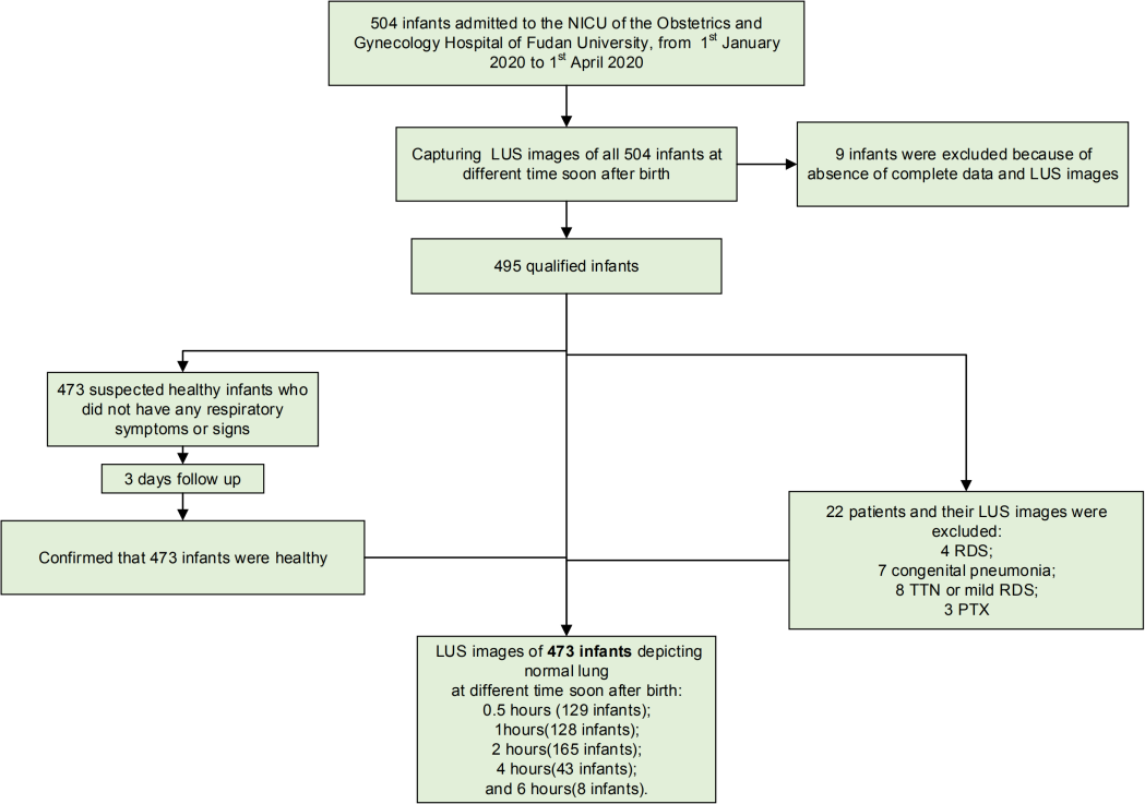
Abstract Background Lung ultrasound (LUS) has been used to diagnose neonatal respiratory diseases. However, few simple method has been reported to predict respiratory support needs(RSN). Our aim was to determine the diagnostic accuracy of a semiquantitative LUS assessment method predicting respiratory support need. Methods We conducted a prospective diagnostic accuracy study following the STARD (Standards for the Reporting of Diagnostic Accuracy Studies) guidelines at a tertiary level academic hospital between 2019 and 2020. After birth, infants were transferred to a monitoring room to determine NICU treatment need. 310 late preterm and term infants with respiratory symptoms enrolled. The LUS assessment was performed for each participant at one of the following times: 0.5 h, 1 h, 2 h, 4 h, and 6 h after birth. Reliability was tested by ROC analysis. Surfactant administration and other RSNs were based on the 2019 European guidelines as well as the infant’s clinical condition. Results 74 have RSN, and 236 were healthy according to a 3-day follow-up confirmation. Six LUS imaging patterns were found. Two “high-risk” patterns were highly correlated with RSN(area under the curve (AUC) = 0.95; 95% CI, 0.92-0.98, p<0.001). This accuracy is supported by the AUC of “low-risk” patterns (0.89, 95% CI, 0.85-0.93, p<0.001). The predictive value of LUS is greater than that of only using respiratory symptoms (e.g., respiratory rate) (AUC of LUS vs AUC of respiratory rate, p<0.01). Conclusions LUS is a useful tool to predict RSN and is more reliable than assessments based on respiratory symptoms alone.

Guannan Xi

and 6 more

Abstract Purpose To distinguish healthy infants from potential patients right after birth using lungultrasound(LUS). Design, Setting and Patients This is a nested case-control study containing 22 lung diseases patients and 473 healthy infants from a total of 504 successive infants. They were admitted to Obstetrics & Gynecology Hospital of Fudan University, Shanghai, China, from 1st January 2020 to 1st April 2020. A newly designed scanning protocol was used to capture LUS images. Sensitivity, specificity, PPV and NPV for predicting healthy infants and patients were calculated individually. The transition process image patterns and thier variation were shown. The relationship between clinic signs and the high-risk image patterns was calculated by Kendall’s tau-b test. Measurements and main results LUS images were captured and its preditive value has been caculated. Four low-risk patterns almost only can be seen in healthy infants(specificity=86.4%, PPV=99.0%) whereas four high-risk patterns can be seen both in healthy infants and patients(specificity=62.4%, PPV=9.6%). High-risk patterns are more likely to be pathological when appearing at oxter and lower back but to be a physiological sign when appearing at Lower and upper of the prothorax. These high-risk patterns are significantly related to clinic sign. All these patterns are alsmost consistent during 6 hours after birth. Conclusions LUS is valid to differentiate healthy infants from potential patients who with mild respiratory difficulty. Four low-risk patterns have high value to predict healthy infants, but four high-risk patterns are not specific enough to discover patients. This criterion is valid for this 6 hours stage.

Guannan Xi

and 6 more

Abstract Purpose To distinguish healthy infants from potential lung disease patients immediately after birth using lung ultrasound (LUS). Design, Setting and Patients This is a nested case-control study containing 22 lung disease patients and 473 healthy infants from a total of 504 consecutive infants. The infants were admitted to the Obstetrics & Gynecology Hospital of Fudan University, Shanghai, China, from 1 January 2020 to 1 April 2020. A newly designed scanning protocol was used to capture LUS images. The sensitivity, specificity, PPV and NPV for predicting healthy infants and patients were calculated individually. The transition process image patterns and their variations are shown. The relationship between clinical signs and high-risk image patterns was calculated by Kendall’s tau-b test. Measurements and main results LUS images were captured, and their predictive value was calculated. Four low-risk patterns could typically be seen only in healthy infants (specificity=86.4%, PPV=99.0%), whereas four high-risk patterns could be seen in both healthy infants and patients (specificity=62.4%, PPV=9.6%). High-risk patterns were more likely to be pathological signs when appearing at the oxter and lower back and physiological signs when appearing at the prothorax. These high-risk patterns are significantly related to clinical signs. All these patterns are consistent during the first 6 hours after birth. Conclusions LUS is a valid modality for differentiating healthy infants from potential patients with mild respiratory difficulty. Four low-risk patterns had high value in predicting healthy infants, but four high-risk patterns were not specific enough to discover lung disease patients.