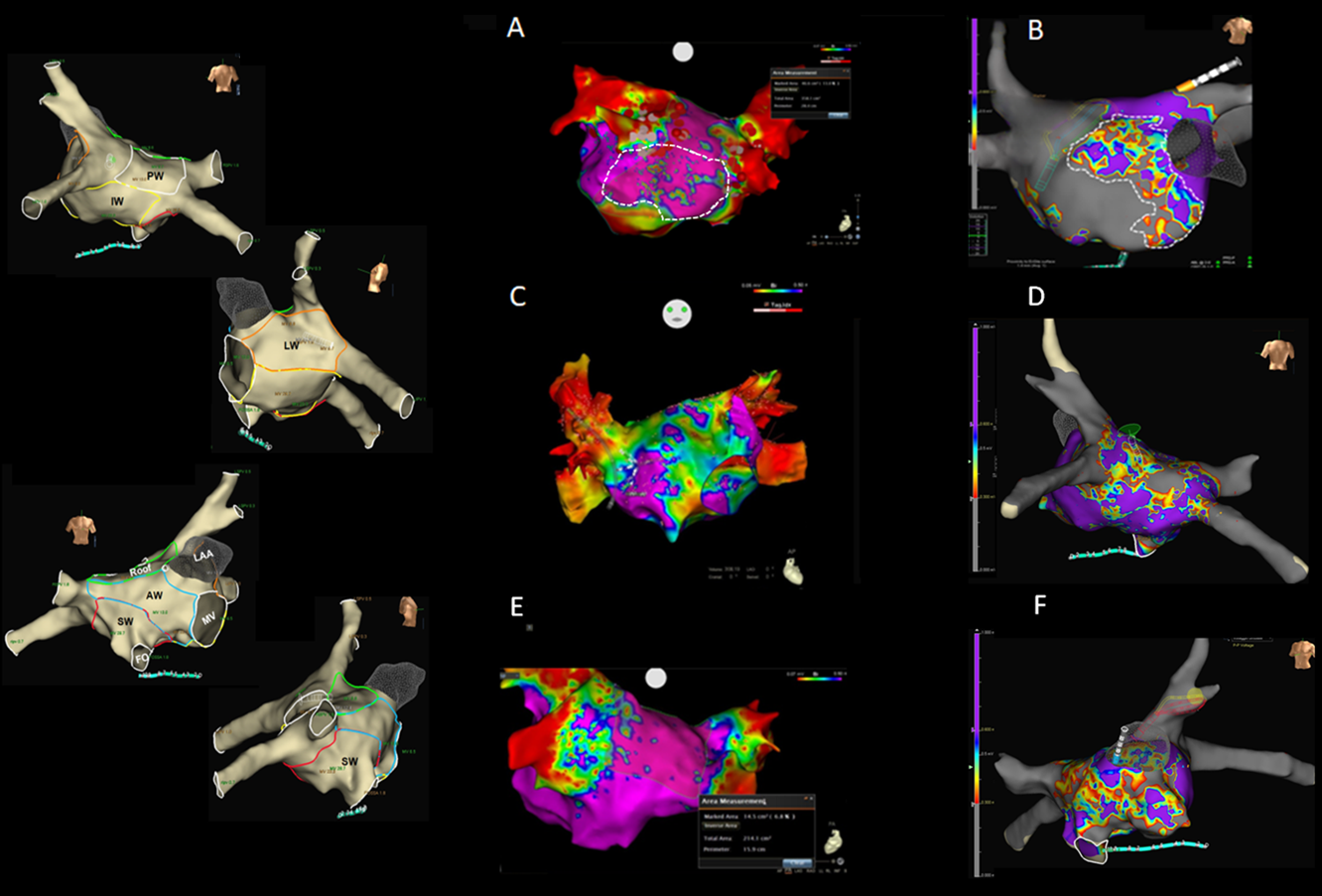
Background. Substrate analysis of the left atrium in patients undergoing atrial fibrillation ablation has limitations when performed by means of simple bipolar acquisition. Objective. To evaluate the incidence of low-voltages (LV) through maps constructed by means of various catheters:multipoltar (MC), omnipolar (OC) and circular catheters (CMC) with the 3D electro-anatomical systems (3d-S) CARTO3 and Ensite-Precision. Methods. To assess LV we acquired maps by means of CMC and MC in the voltage range 0.05-0.5 mV in 70 patients in sinus rhythm. In case of OC only, we made an intra-patient comparison of bipolar maps constructed in along, across and HD-Wave configurations by means of Ensite-Precision in the ranges of 0.05-0.5 mV and 0.5-1.0 mV. Basing on this comparison, we chose the range that best identified LV and characterized patchy fibrosis by analyzing a set of different colors (qualitative analysis). Finally, we performed a quantitative analysis of LV by applying the qualitative characteristics described above. Results. Basing on our settings, the optimal range for OC was 0.3-0.6 mV. OC revealed smaller LV areas than MC (p <0.05 or p <0.001), except in the lateral wall. No significant differences were observed between CMCs. The same rates of AF recurrence were found for OC and MC during the follow-up period. Conclusions.In our experience, OC does not present the limits of bipolar HD maps, though further studies are needed in order to confirm that 0.3-0.6 mV as LV optimal voltage range.