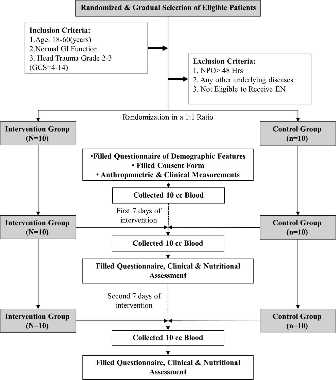
Aim: In Traumatic brain injury (TBI) patients, a complex cascade of inflammatory responses is frequently observed following trauma. Numerous dietary agents have long been found to have potential in modulating inflammatory responses. This pilot study, designed an enteral formula with low inflammatory properties based on the dietary inflammatory index (DII®) and evaluated its effect on inflammatory and metabolic factors in critically ill TBI patients. Methods: This Single-blind randomized controlled pilot study conducted at the Neurosurgical ICU of Shahid Kamyab Hospital (Mashhad, Iran). A total of 20 TBI patients were randomly assigned to receive either low-DII-score or standard formula at the Intensive Care Unit (ICU). The primary outcomes of the study included clinical status, inflammatory biomarkers, APACHE II, SAPS II, SOFA, and NUTRIC scores. Results: The trial groups did not differ significantly on baseline values. Following 14 days of intervention, there was a statistically significant decrease in the APACHE II, SAPS II, and NUTRIC scores and a significant increase in GCS score in the low-DII-score formula group compared to the standard formula group. Over two weeks, high sensitivity c-reactive protein (hs-CRP) values -2.73 (95% CI: -3.67, -1.79) mg/dL in the low-DII-score formula group vs. 0.65 (95% CI: -0.29, 1.58) mg/dL in controls. Moreover, the length of hospital stay was longer for the standard formula group than for the low-DII-score formula group. Conclusion: The low-DII-score formula improves inflammatory factors (serum hs-CRP) and metabolic biomarkers (LDL-c and FBS). Furthermore, clinical outcomes, including the length of hospital stay and disease severity appear to be enhanced.