Duygu Temiz Karadag

and 15 more

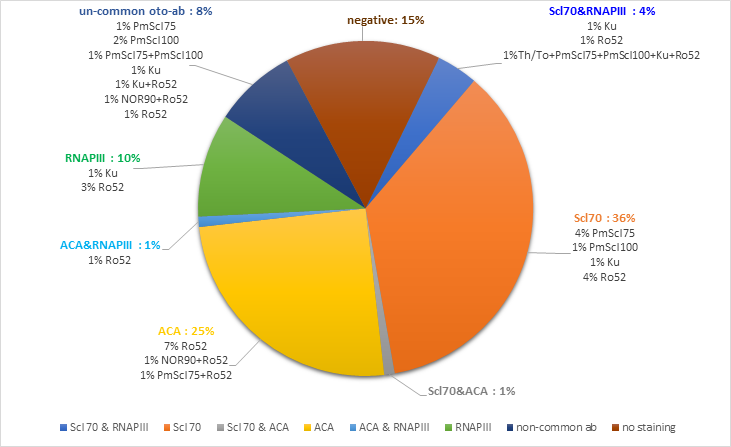
Background/aim: To investigate the frequency and clinical relevance of an extended autoantibody (ab) profile in patients with SSc. Materials and Methods: In this cross-sectional study, serum from 100 consecutive patients was subjected to indirect immunofluorescence (HEp-20-10/primate liver mosaic) and Systemic Sclerosis Profile by EUROIMMUN (Lübeck, Germany) to evaluate ANA and autoantibodies against 13 different autoantibodies in patients with SSc less than three years. Results: 93 of 100 patients were positive for ANA by indirect immunofluorescence (IIF). The prevalence of Anti-Scl70 ab was 41%, anti-centromere (ACA) 27%, and anti-RNA polymerase (RNAPIII) 15%. Scl70 was more associated with diffuse subtype (p<0.001), ILD (p<0.001), and high mRSS (p=0.002); ACA with limited disease (p<0.001), less ILD (p<0.001), overlap (p=0.017) and low mRSS (p=0.024); RNAPIII with diffuse disease (p=0.027), ILD (p=0.016) and high mRSS (p=0.001). Fifty-three patients showed single positivity (26 anti-Scl70, 16 ACA, 6 anti-RNAPIII, 1 anti-Ku ab, 1 anti-PM/Scl100, 2 anti-PM/Scl75, 1 anti-Ro52), whereas 32 patients had multiple auto-antibody positivities. Among common SSc-specific autoantibodies, Scl70 and RNAPIII showed the highest co-occurrence (n=4). One patient was simultaneously positive for anti-RNAPIII ab and ACA, and one was positive for ACA and Scl70. The clinical features were not statistically different between single and multiple autoantibody-positivity for common SSc-specific autoantibodies (ACA, Scl70, and RNAP III), except for digital ulcer in multi-antibody positive ACA group (p=0.019). Conclusion: Based on our results, co-expression of auto-antibodies is not uncommon in SSc patients. Although SSc-specific auto-antibodies generally show known clinical features, the clinical presentation of the co-expression in specific and non-specific auto-antibody positivity continues to be important.

Duygu Temiz Karadag

and 4 more

Objectives: To investigate lung volume and density in patients with SSc and changes in these parameters due to PF, using a software-aided image quantification method, and compare this with a matched healthy control group. Methods: Thoracic high-resolution computed tomography (HRCT) images of patients and controls were analyzed using Myrian XP Lung 3D software. Right, and left lung densities and volumes were calculated separately by a blinded operator. Results were analyzed between subgroups to investigate associations with the clinical features. Results: A total of 135 patients with SSc and 38 healthy controls (HC) were included. Characteristics of the SSc patients were 94 (69.6%) without PF, 85.4% female, mean age 49.8 (15.4) years; 41 (30.4%) with PF, 88.3% female, mean age 50.2 (11.5) years and HC group were 89.5% Female, mean age 52.2 (5.8) years. The right and left lung densities were significantly higher, and right and left lung volumes were significantly lower in the SSc patients with signs of fibrosis than those without and HC (p<0.001 and p<0.001; p=0.006 and p=0.002, respectively). After excluding patients with PF, right and left lung densities and volumes differed significantly between diffuse cutaneous SSc, limited cutaneous SSc, and HC (p=0.002 and p<0.001; p=0.045 and p=0.044, respectively). Patients who developed PF during follow-up had significantly lower baseline right and left lung densities than those who did not (p=0.018; p=0.014, respectively). Forced vital capacity and carbon monoxide diffusing capacity showed weak correlation with lung densities and volumes in patients without PF and moderate to high correlation in PF patients. Conclusion: Lung density and volume in SSc patients changed significantly in those with PF and those without. Quantitative information extracted by soft-ware aided methods may contribute more to the detection, screening, and risk prediction in SSc related PF.