Chuan Wang

and 9 more

Half of patients with heart failure are presented with preserved ejection fraction (HFpEF). The pathophysiology of these patients is complex but increased left ventricular (LV) stiffness is proved to play a key role. However, the application of this parameter was limited since its measurements requires invasive catheterization. With advances in ultrasound technology, new advances have been achieved in the assessment of LV chamber or myocardial stiffness using noninvasive echocardiography. Therefore, this review was carried out to summarize the pathophysiological mechanisms, correlations with invasive LV stiffness constant, applications in different populations as well as the limitations of echocardiography-derived indices for assessment of both LV chamber and myocardial stiffness. LV chamber stiffness indices such as E/e’/LVEDV, E/SRe/LVEDV, and DPVQ were derived on the basis of the relationship between echocardiographic parameters of LVFP and LV size. However, all these methods are surrogate and lumped measurements, relying on E/e’ or E/SRe for evaluating LVFP. The limitations of E/e’ or E/SRe in assessment of LVFP may contribute to the moderate correlation between E/e’/LVEDV or E/SRe/LVEDV and LV stiffness constant. Even the best validated measurement (DPVQ) is considered unreliable in the individual patient. Compared to E/e’/LVEDV and E/SRe/LVEDV, I PVA/IA and F PVA/FA may display better performance in assessing LV chamber stiffness as evidenced by a higher correlation with LV stiffness constant. However, only one study has been conducted in the literature on the exploration and application of I PVA/I A and F PVA/F A, and its accuracy in assessing LV chamber stiffness remains to be confirmed. In terms of echocardiographic indices for LV myocardial stiffness evaluation, the parameters of EMI/DWS, iVP and SWI were proposed. Despite alteration of DWS and its predictive value of adverse outcomes in various populations have been widely validated, it was found that DWS may be better considered as an overall marker of cardiac function performance instead of pure myocardial stiffness. As for the iVP and SWI, the validities of these two indices in assessing LV myocardial stiffness have not been confirmed in invasive studies. More echocardiographic indices with higher sensitivities and specificities warrant to be further uncovered to evaluate LV stiffness.

Shuran Shao

and 10 more

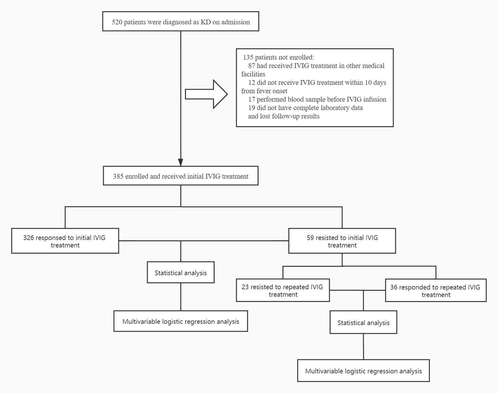
Background: Intravenous immunoglobulin (IVIG) resistance prediction remains substantial in Kawasaki disease (KD), with limited data on the predictive value of coagulation profile for IVIG resistance, particularly for repeated IVIG resistance. Therefore, the aim of our study was to testify the predictive validity of coagulation profile for both initial and repeated IVIG resistance in KD. Methods: A total of 385 KD patients were prospectively recruited between April in 2015 and May in 2019. Coagulation and other profiles were evaluated between IVIG-responsive and IVIG-resistant groups. Multivariate logistic regression analysis was applied to determine the association between coagulation profiles and IVIG resistance. ROC curves analysis was further performed to assess validity of coagulation profiles in predicting both initial and repeated IVIG resistance. Results: PT, APTT and D-dimer were significantly increased in initial IVIG-resistant group with ATIII significantly reduced. Meanwhile, ATIII was declined markedly in repeated IVIG-resistant patients. PT, APTT, D-dimer and ATIII cutoff values of 13.95 s, 41.15 s, 1.48 mg/l, and 89.5% yielded sensitivities of 73%, 32%, 71%, 81%; specificities of 55%, 88%, 62%, 51% for predicting initial IVIG resistance, respectively. The cutoff value of ATIII for predicting repeated IVIG resistance was 68.5%, with sensitivity of 71% and specificity of 55%. Multivariate logistic regression analysis showed that PT, APTT, D-dimer and ATIII were independent risk factors for initial IVIG resistant patients with KD. Conclusions: Coagulation profiles were significantly dysregulated in KD patients. Some of them particularly ATIII may serve as complementary laboratory markers for prediction of both initial and repeated IVIG resistance.