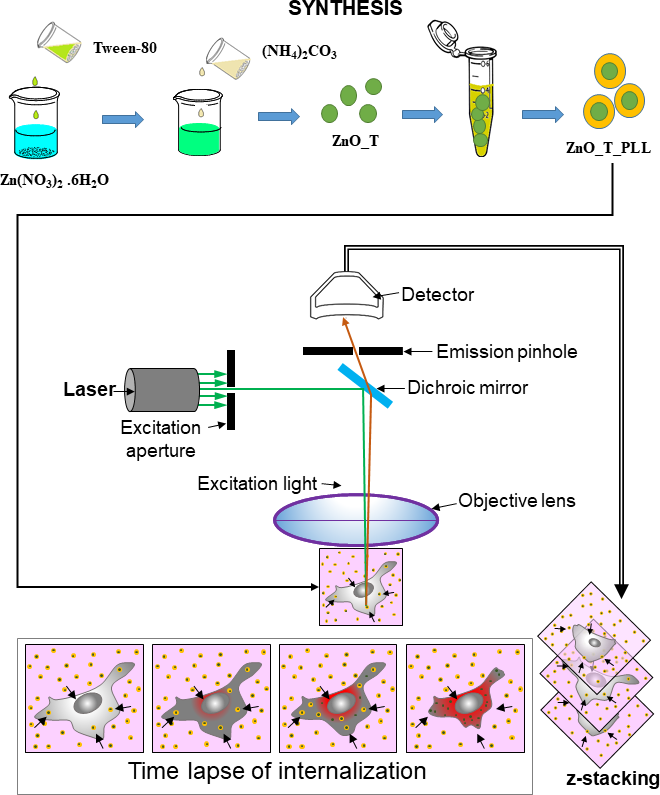
Abstract Generally, investigations on nanomedicine involve conventional imaging techniques for obtaining static images on nanoparticle internalization at a single time point where various phases can be overlooked. In contrast, 3D live-cell imaging can be used for obtaining cellular retention of drugs at various phases, and cells can be followed for days. This article demonstrates the application of time-lapse microscopy in the investigation of Poly-L-lysine coated ZnO nanoparticle dynamics. In this work, a laser scanning confocal microscope has been employed to quantify the dynamics of internalization particles and reactive oxygen species generation (ROS) using volumetric imaging. Firstly, we show that simultaneous spatial mapping of nanoparticle uptake in MCF-7 cells and ROS in a single cell can be used to identify the interdependence between the accumulation of particles and ROS generation. Secondly, monitoring of ROS formation and cytotoxicity using the same imaging platform offers an advantage over monitoring these parameters using various instruments. Finally, the ability of the fluorescent particles in inducing a significant reduction in cell viability suggests its potential to be used as a therapeutic agent. The proposed framework opens up a new avenue of research for investigating mechanistic aspects of ZnO particle adsorption in vitro through long term imaging. Keywords: Fluorescent ZnO particle, Time-lapse microscopy, 3D Live-cell imaging, laser scanning confocal microscope, Reactive oxygen species