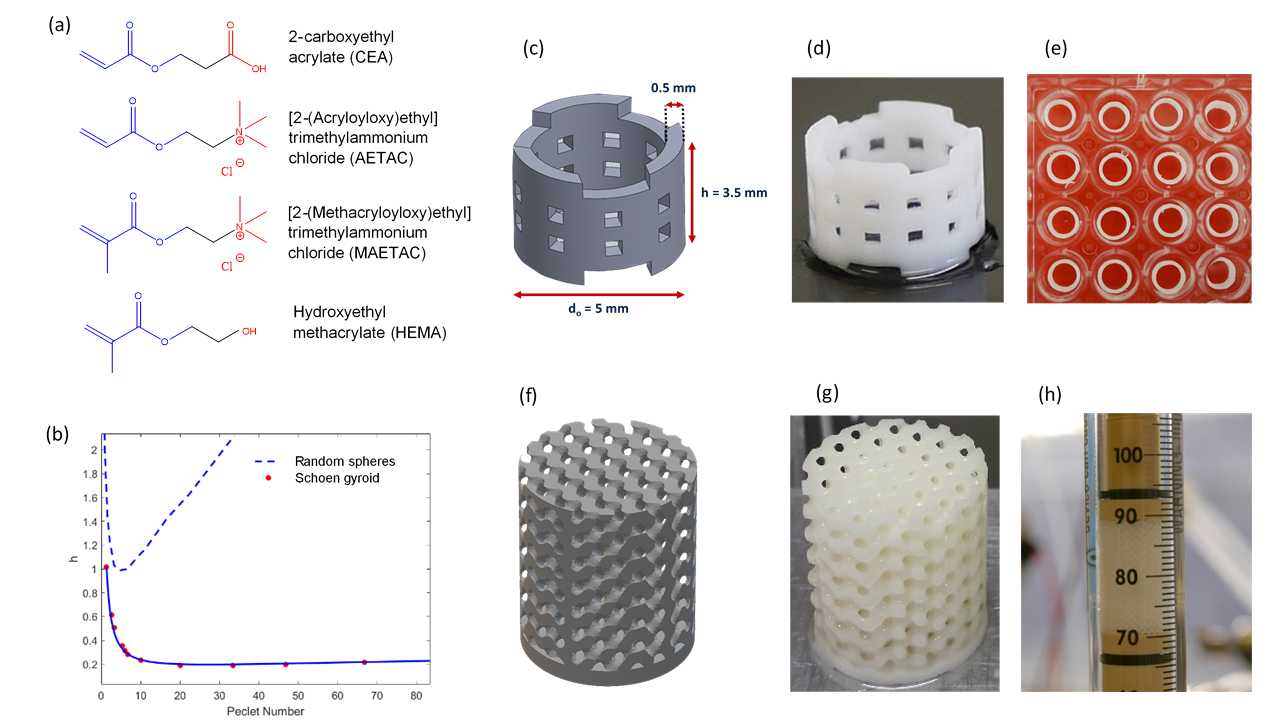
3D printing is revolutionizing many industrial sectors and has the potential to enhance also the biotechnology and bioprocessing fields. Here, we propose a new flexible material formulation to 3D print support matrices with complex, perfectly ordered morphology and with tuneable properties to suit a range of applications in bioprocess engineering. Supports for packed-bed operations were fabricated using functional monomers as the key ingredients, enabling matrices with bespoke chemistry such as charged groups, chemical moieties for further functionalization, and hydrophobic/hydrophilic groups. Other ingredients, e.g. crosslinkers and porogens, provide the opportunity to further tune the mechanical properties of the supports and the morphology of their porous network. Through this approach, we fabricated and demonstrated the operation of Schoen gyroid columns with I) positive and negative charges for ion-exchange chromatography, II) enzyme bioreactors with immobilized trypsin to catalyse hydrolysis, and III) bacterial biofilms bioreactors for fuel desulfurization. We expect this approach will enable simple, cost-effective and flexible fabrication of customized supports in biotechnology and bioengineering.