Milena Leo

and 11 more

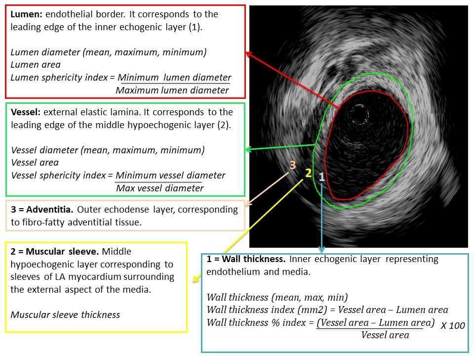
Introduction. Preliminary data in human suggest that both Intracardiac echocardiography (ICE) and Intravascular ultrasound (IVUS) can be used for real-time information on the left atrial (LA) wall thickness and on the acute tissue changes produced by energy delivery. This pilot study was conducted to compare ICE and IVUS for real-time LA wall imaging and assessment of acute tissue changes produced by radiofrequency (RF), cryo and laser catheter ablation. Methods Patients scheduled for RF, cryoballoon or laser balloon Pulmonary Vein Isolation (PVI) catheter ablation were enrolled. Each pulmonary vein (PV) was imaged immediately before and after ablation with either ICE or IVUS. The performance of ICE and IVUS for imaging were compared. Pre- and post-ablation measurements (lumen and vessel diameters, areas and sphericity indexes, wall thickness and muscular sleeve thickness) were taken at the level of each PV ostium. Results A total of 48 PVs in 12 patients were imaged before and after ablation. Compared to IVUS, ICE showed higher imaging quality and inter-observer reproducibility of the PV measurements obtained. Acute wall thickening suggestive of oedema was observed after RF treatment (p = 0.003) and laser treatment (p = 0.003) but not after cryoablation (p = 0.69). Conclusions Our pilot study suggests that ICE is preferable to IVUS for LA wall thickness imaging at the LA-PV junctions during ablation. Ablation causes acute wall thickening when using RF or laser energy, but not cryoenergy delivery. Larger studies are needed to confirm these preliminary findings.

Michael Pope

and 6 more

Background Charge density mapping of atrial fibrillation (AF) reveals dynamic patterns of localised rotational activation (LRA), irregular activation (LIA) and focal firing (FF). Their spatial stability, conduction characteristics and the optimal duration of mapping required to reveal these phenomena and has not been explored. Methods Bi-atrial mapping of AF propagation was undertaken and variability of activation patterns quantified up to a duration of 30-seconds(s). The frequency of each pattern was quantified at each vertex of the chamber over 2 separate 30s recordings prior to ablation and R2 calculated to quantify spatial stability. Regions with the highest frequency were identified at increasing time durations and compared to the result over 30s using Cohen’s kappa. Properties of regions with the most stable patterns were assessed during sinus rhythm and extrastimulus pacing. Results In twenty-one patients, 62 paired LA and RA maps were obtained. LIA was highly spatially stable with R2 between maps of 0.83(0.71-0.88) compared to 0.39(0.24-0.57) and 0.64(0.54-0.73) for LRA and FF, respectively. LIA was also most temporally stable with a kappa of >0.8 reached by 12s. LRA showed greatest variability with kappa>0.8 only after 22s. Regions of LIA were of normal voltage amplitude (1.09mv) but showed increased conduction heterogeneity during extrastimulus pacing (p=0.0480). Conclusion Irregular activation patterns characterised by changing wavefront direction are temporally and spatially stable in contrast with rotational patterns that are transient with least spatial stability. Focal activation appears of intermediate stability. Regions of LIA show increased heterogeneity following extrastimulus pacing and may represent fixed anatomical substrate.