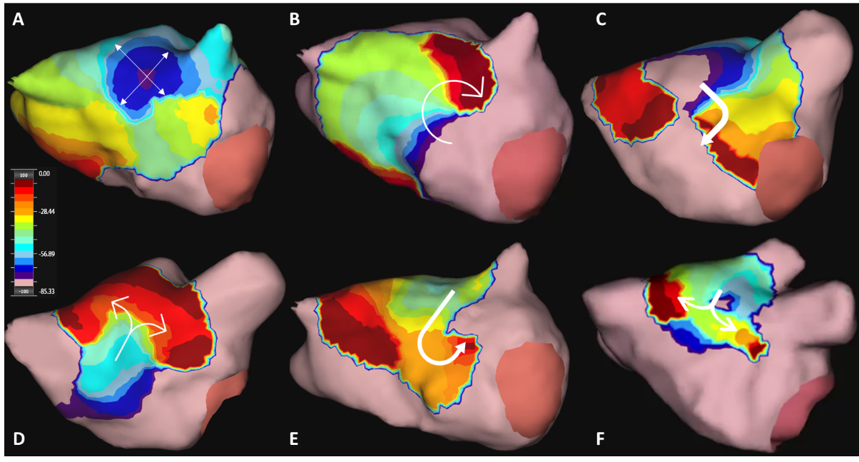
Background Charge density mapping of atrial fibrillation (AF) reveals dynamic patterns of localised rotational activation (LRA), irregular activation (LIA) and focal firing (FF). Their spatial stability, conduction characteristics and the optimal duration of mapping required to reveal these phenomena and has not been explored. Methods Bi-atrial mapping of AF propagation was undertaken and variability of activation patterns quantified up to a duration of 30-seconds(s). The frequency of each pattern was quantified at each vertex of the chamber over 2 separate 30s recordings prior to ablation and R2 calculated to quantify spatial stability. Regions with the highest frequency were identified at increasing time durations and compared to the result over 30s using Cohen’s kappa. Properties of regions with the most stable patterns were assessed during sinus rhythm and extrastimulus pacing. Results In twenty-one patients, 62 paired LA and RA maps were obtained. LIA was highly spatially stable with R2 between maps of 0.83(0.71-0.88) compared to 0.39(0.24-0.57) and 0.64(0.54-0.73) for LRA and FF, respectively. LIA was also most temporally stable with a kappa of >0.8 reached by 12s. LRA showed greatest variability with kappa>0.8 only after 22s. Regions of LIA were of normal voltage amplitude (1.09mv) but showed increased conduction heterogeneity during extrastimulus pacing (p=0.0480). Conclusion Irregular activation patterns characterised by changing wavefront direction are temporally and spatially stable in contrast with rotational patterns that are transient with least spatial stability. Focal activation appears of intermediate stability. Regions of LIA show increased heterogeneity following extrastimulus pacing and may represent fixed anatomical substrate.