Dong Geum Shin

and 6 more

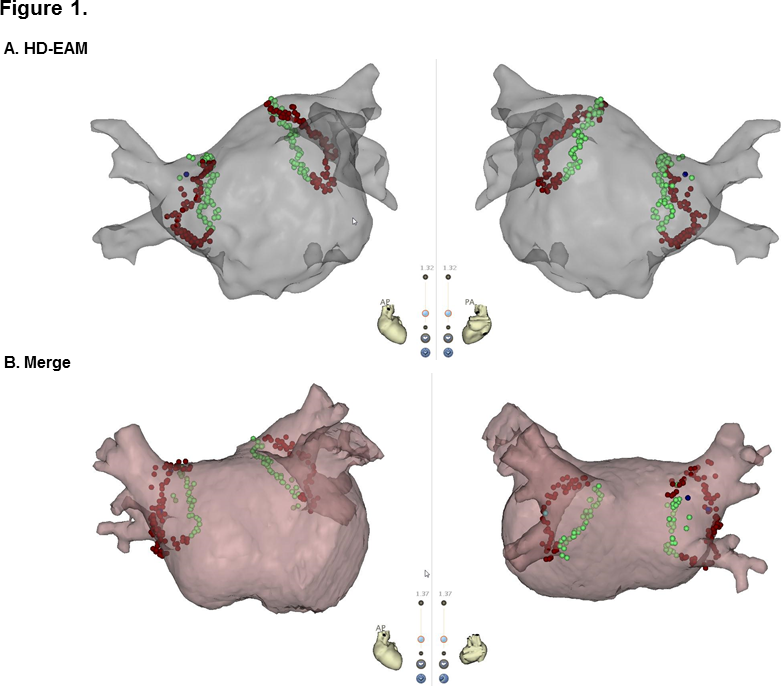
Introduction: Integration of electroanatomical map (EAM) with preacquired three-dimensional (3D) cardiac images provides detailed appreciation of the complex anatomy of the left atrium (LA) and pulmonary vein (PV). High-density (HD) multi-electrode mapping catheters have enabled creating more accurate EAM reflecting real-time volume-rendered LA-PV geometry during atrial fibrillation (AF) ablation. However, no study has compared the outcomes of AF ablation using HD-EAM versus 3D-merged map. We aimed to investigate the procedural and clinical outcomes of AF ablation with HD-EAM (HD-EAM group) versus 3D-merged map (Merge group). Methods: One hundred patients (59.5±11.5years, 53% with paroxysmal AF [PAF]) were randomly assigned (1:1) to HD-EAM or Merged group. HD multi-electrode mapping and contact force (CF)-sensing catheters were used to create virtual LA-PV chamber and to perform wide antral circumferential ablation (WACA), respectively. Results: The two groups showed no significant differences in baseline characteristics and procedural data including ablation time, fluoroscopy time, LA voltage, and CF. PV isolation with a single WACA line was achieved in 21 (42%) and 27 (54%) patients in the Merge and HD-EAM groups, respectively (P=NS). CF was significantly lower in lesions with gap than lesions without gap after a single WACA (7.3±7.3 g vs. 16.0±8.3, respectively, P<0.001). During the 12-month follow-up, no significant difference in AF recurrence was observed between two groups, irrespective of AF type. In multivariate analysis, non-PAF was an independent risk factor for AF recurrence. Conclusion: Integration of 3D cardiac imaging did not improve procedural and clinical outcomes. HD-EAM provides an accurate real-time LA geometry.

Dong Geum Shin

and 3 more

Introduction: Single-shot ablation has emerged as an effective technique for index atrial fibrillation (AF) ablation, with an advantage of short procedure time. Although recent guidelines recommend peri-procedural uninterrupted oral anticoagulants (OACs), the intra-procedural anticoagulation strategy remains uncertain under non-vitamin K OACs (NOACs). We investigated procedural safety of a single bolus administration of heparin without activated clotting time (ACT) measurement during cryoballoon ablation (CBA). Methods: Two hundred patients (64.2±10.0years, 70% with non-paroxysmal AF) who underwent CBA with uninterrupted NOACs were randomly assigned to No-ACT group and ACT group. A bolus of heparin (100 U/kg) was routinely administered immediately after transseptal puncture. In the ACT group, an additional injection of heparin (30 U/kg) was administered if ACT at 30-min after the initial bolus was <300 s. Results: There were no differences in baseline characteristics including CHA2DS2-VASc score between two groups. The left atrium indwelling and procedure times were 60.4±13.1 min and 78.9±13.9 min, respectively and not significantly different between two groups. The mean ACT was 335.2±59.9 s in the ACT group. Any bleeding rate was 3.2% in all patients and there was no statistically difference in bleeding complications between two groups. In the ACT group, groin hematoma, laryngopharyngeal bleeding, and hemoptysis occurred in 3, 1, and 1 patient, respectively. Cardiac tamponade occurred in 1 patient in the No-ACT group. No thromboembolic events occurred during the 30-day follow-up after CBA. Conclusion: Single bolus administration of heparin without ACT measurement is a feasible anticoagulation strategy for CBA in patients with uninterrupted NOACs intake.