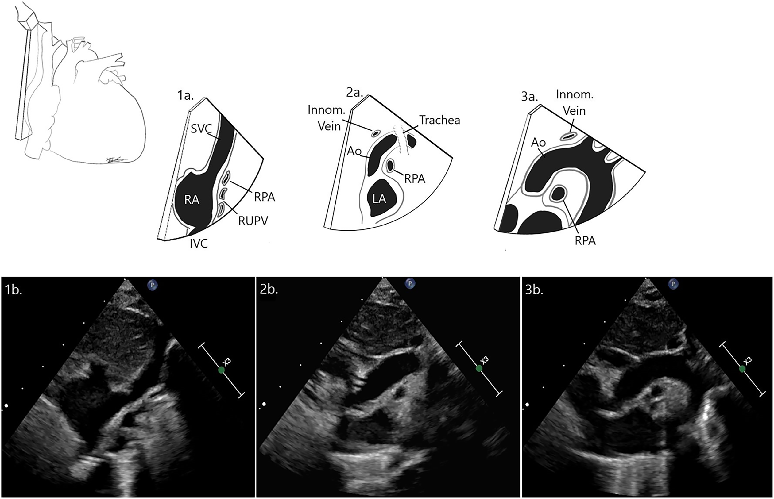
Objective: Diagnosis of Aortic arch (AoA) anatomy is critical for planning cardiac surgery/intervention and in diagnosing associated congenital heart defects. AoA sidedness is traditionally diagnosed with echocardiography as being contralateral to the direction of the first brachiocephalic artery in suprasternal view, but this method can be challenged by numerous anatomic variants and clinical conditions. The objective of this study was to assess feasibility of trachea visualization with echocardiography in pediatric patients, and using this landmark to identify AoA sidedness and potential for double aortic arch (DAA). Methods: A prospective study was performed on patients <18 years old who were undergoing Chest CT/MRI to serve as gold standard for confirming AoA anatomy. A right-to-left echocardiographic sagittal sweep was performed from the suprasternal notch and used to categorize 1) Left AoA = right SVC-trachea-AoA, 2) Right AoA= SVC-AoA-trachea, 3) DAA = SVC-AoA-trachea-AoA. The proportion of successful sweeps and diagnostic accuracy were calculated. Results: 100 consecutive patients were scanned (44% female; median age of 8.8 yr, range 2d–17.9 yr; median BSA 1.14 m 2, range 0.2–2.7; right AOA in 4%). Diagnosis of AoA sidedness was possible in 97% (95% CI: 94–100%, p < 0.01) and correct in 100% when the trachea was seen. Conclusion: Tracheal imaging with echo is reliable, easy, and reproducible method in patients of various sizes and levels of acuity to define AoA sidedness.